Hadatomo™ Photoacoustic Microscope
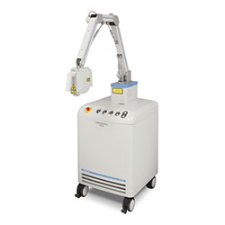
Noninvasive, High Contrast Imaging of Blood Vessels
The new Photoacoustic Microscope Hadatomo™ WEL5100 enables imaging of blood vessels within the dermis to a depth of 3mm. The differing absorptive characteristics of hemoglobin and tissue allow the target area to be selectively imaged in high contrast.
Suggested Applications
- Analyze progress of skin grafts in plastic surgery procedures
- Check regrowth of blood vessels in cultured tissues in regenerative medicine procedures
- Imaging of Blood Vessels to a Depth of 3mm
The principle of Photoacoustic Imaging
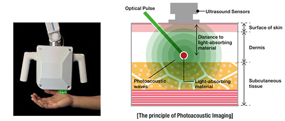
Advantest's new photoacoustic microscope combines the propagation characteristics of ultrasound and the absorption characteristics of light into a new hybrid imaging method. By using ultrasound technology, it can obtain accurate information to a depth of several millimeters: Hemoglobin selectively absorbs the energy of pulsed light and returns ultrasonic waves to the surface of the skin, where they can be captured by sensors. Based on how long it takes for the waves to return, the depth of the target can be accurately measured and imaged.
WEL 5100 Imaging Examples (1)
Case study: area of the palm
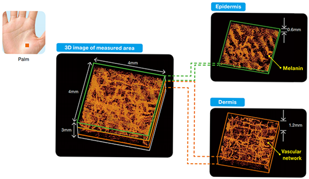
3D image of a 4mm x 4mm x 3mm area of the palm, including the epidermis and dermis.
It was further analyzed into epidermis (0.6 mm) and dermis (1.2 mm).
WEL 5100 Imaging Examples (2)
Case study:measurement of blood circulation change in forearm after application of pressure
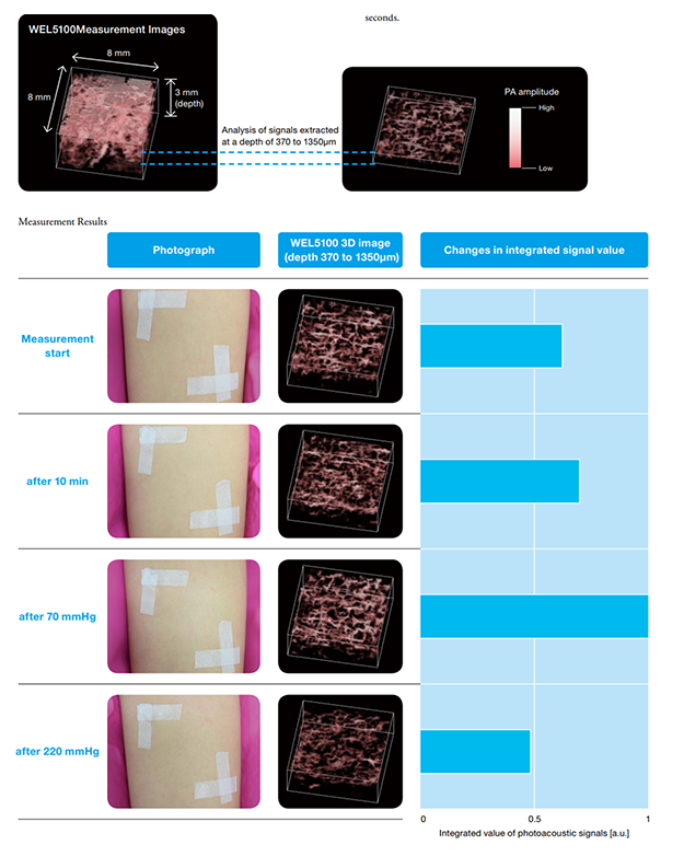
We used the WEL 5100 to measure Photoacoustic(PA) signals originating from blood previous to applying pressure with a cuff, and 10 minutes afterwards. The cuff applied 70 mmHg pressure for 30 seconds, and 220 mmHg pressure for 30 seconds.
Examples using the WEL5100
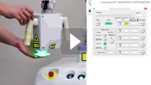
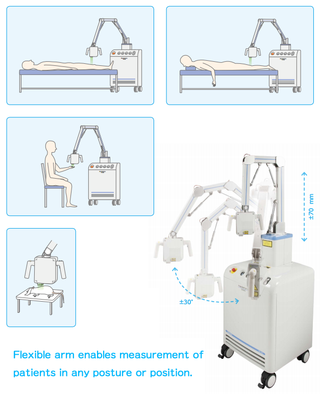
Operation of the WEL5100
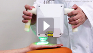
The WEL5100 features easy operabillity and a flexible arm that enables measurement of object in any pisture or position.
Exampe application - part1 (Phantom)
Hadatomo™ enables imaging of tissue within the object under test to a depth of 3mm.
Product Specifications
| Model Name | WEL5100 |
|---|---|
| Wavelength | 532nm |
| Pulse Length | < 2 ns |
| Optical Energy | < 100 μJ/pulse* |
| Measurement Time | 20 s/40 s/80 s/160 s |
| Measurement Area | 4 x 4 x 3(depth)mm/ 8 x 8 x 3(depth)mm |
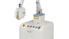
| Dimensions | Approx. 540(w) x 545(D) x 1546(H) mm |
| Weight | < 110kg |
*Optical(irradiation) energy is constant, but repetition frequency may vary depending on measurement conditions.
Manufacturer:ADVANTEST CORPORATION
Saitama R&D Center 1-5, Shin-tone, Kazo-shi
Saitama 349-1158
Selling agency:KYODO INTERNATIONAL INC.
検索用入力欄
記入時注意事項
- テーブルの行列は変更しないでください。
- 分類が複数ある場合は、| ←半角の縦棒で区切って連続で入力してください。
また、区切り文字の前後に空白は入れないでください。
| キーワード分類1 | Tissue |
|---|---|
| キーワード分類2 | Imaging |
| キーワード分類3 |
|
| 製品カテゴリ1 | Device |
| 製品カテゴリ2 | Imaging |
| 分野 | Regenerative medicine|Biological imaging|Pathological animal model |
| サムネイル画像 |
|
| 説明文 | N]on-invasively image blood vessel structure. |
| リダイレクトURL |
|